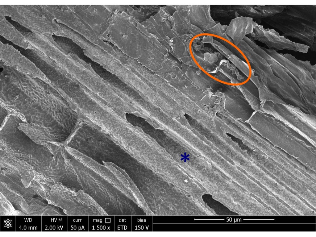
Evidently not. I mounted three of each type, and since I had two other mounted from the initial reconnoiter, that makes five. Friday evening I had 90 min on the scanning electron microscope. I picked a low-ish magnification, one that gave an image corresponding to about two tenths of a millimeter from one side to the other. Then I started at one end of a section and walked toward the other, following the fibers. This is straightforward because the fibers run like a trail up each side of the section, just a few cells in from the epidermis. I took about 10 images per section, and with four sections in play (I didn’t get the original stub with the first ones) that meant forty images from the non-bleached, and another forty from the bleached. The magnification was high enough to spot residual cytoplasm but low enough to let me survey a fair amount of real estate.

The no-bleach ones retained a little bit of cytoplasm on some walls (Figure 1). However the majority of walls were clean. Clean enough that there would be no trouble taking as many high magnification images as desired. I think we can safely omit the bleach. I suppose there is a formal possibility that one of the mutants that is intermediate in severity between the wild type and this most extreme mutant could turn out to hang onto cytoplasm more tightly. Or that preparations made on various days would have cytoplasm with differential stickiness. Both possibilities seem remote. I think we have a green light for a full comparison.