Hi Lab Fab fans. UMass changed blogging platforms which forced me to take a few weeks off while they retooled. Things seem ok now (?). The following post was written for Jan 14th but still holds. By the way if you like Lab Fab, consider subscribing to never miss a post. Its free! Scroll down below the socks and the link-list to add your email and hit the button. Now, on to science…
The past week, I used the two-photon microscope. On last week’s blog post, I explained my reason; finally, I got to try it.
I was nervous walking over the Medical School, with the plate of seedlings nestled in my backpack to shield them from the cold weather. My nervousness increased as I neared the door to the wing housing the imaging suite: People were milling around on the sidewalk. As I drew closer, I heard the loud chirping of an alarm. I could see another throng in front of a distant entrance. A fire? No way to enter. I didn’t have the cell number of Dean, the person who manages the two-photon instrument and who I was meant to be meeting. I waited.
Eventually, the sirens went silent and we all trouped in. I found Dean and despite the loss of 30 min or so we both agreed to have a go. He led me to the room containing the instrument. If I hadn’t known it was a microscope, I am not sure I would have known it was a microscope (Fig. 1). Confocals I have seen or used take a more or less ordinary microscope stand and hang the fancy optical accessories on the sides and back. By contrast, the Olympus1 two-photon2 microscope stand was purpose built and looked like a tank. The only concessions to an ordinary microscope were a pair of eyepieces at the front, which in the event we never even used, and a single objective lens. Apparently, the operation is super-sensitive to stray light.

Figure 1. The Olympus FVMPE-RS Multi-photon. Note, this is not the one at the University of Birmingham Medical, rather it comes from here. But looks fairly similar.
My heart sank because all that enclosing was going to make it challenging to find a way to get the liquid crystals into the beam. Putting that issue aside for later, we turned to the job at hand —checking how the instrument handled the roots stained with Calcofluor. I opened the square Petri dish with the seedlings growing with their roots in agar and placed the dish on the coffee table-sized motorized platform that replaced the typical slide holder for the microscope. We found a root by seeing the scattering when ordinary blue light was allowed to emerge from the objective. Then, thick black curtains were zipped down removing the entire instrument from sight. From then on, everything was under computer control.
We dialed in 700 nm exciting light and let the optics rip. And wow! The root shone like a newly waxed floor on a summer afternoon (Fig. 2). No problem with using 700 nm photons to excite Calcofluor. Likewise, there was no problem imaging with the roots in agar. Test aced!
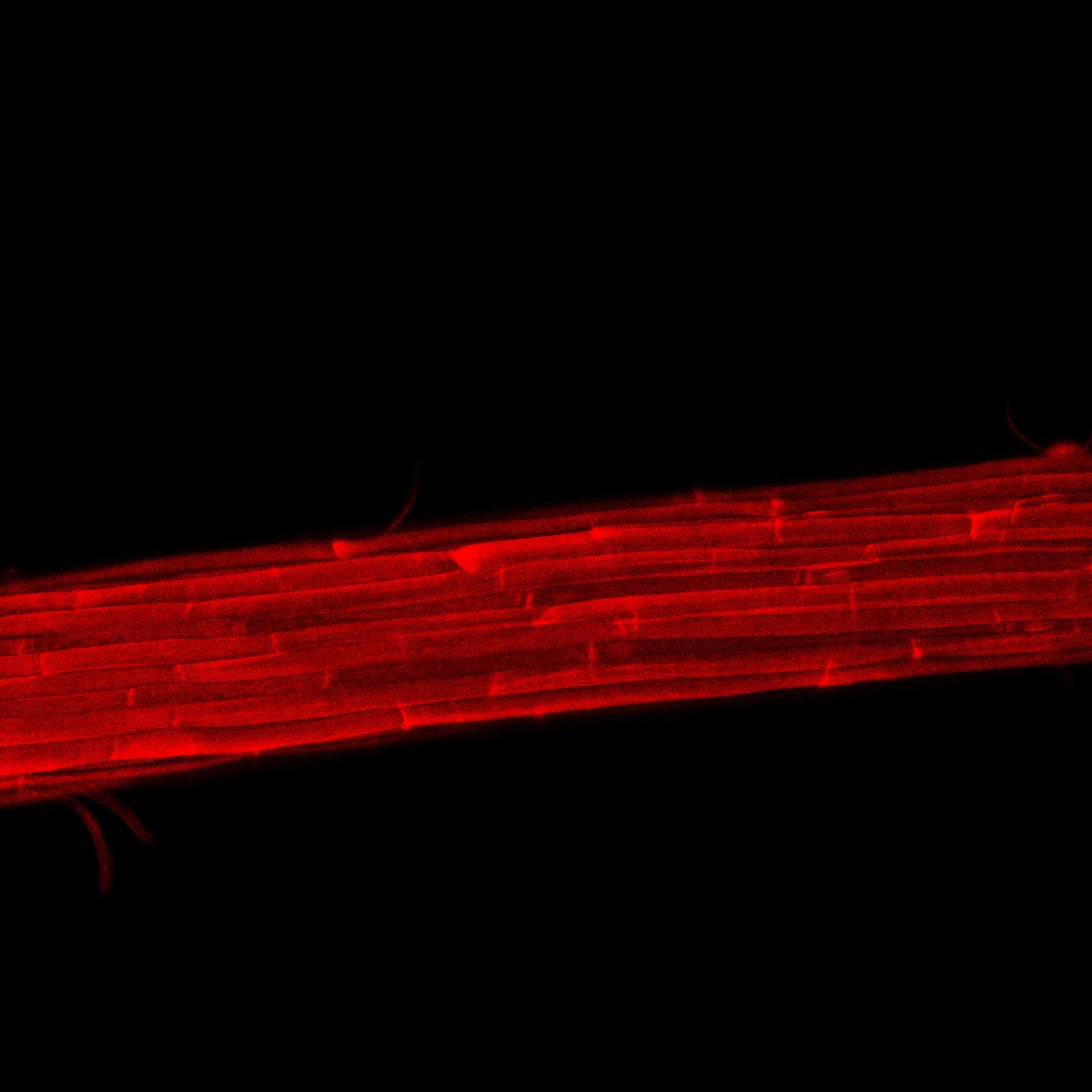
Figure 2. This image of the elongation zone of an arabidopsis is similar to what I got on the two photon. But I don’t have access right now to those images. This image will have to do. The red fluorescence of the cells in face view is the cell wall. Root diameter here is ~100 µm.
All good. But all for naught if the liquid crystals cannot be slotted into the beam. The best place for them is on the excitation side; in that case all that is needed to insert are the pair of liquid crystals (along with the wires for electronic control). The other place is on the emission side. This place has the extra requirements of adding a linear polarizer somewhere after the liquid crystals and, in addition, converting the laser to unpolarized or circular polarization for excitation. As much as it would be great to use two-photon microscopy for this project, I am dubious. Still, Dean is checking with the Olympus engineers. Maybe …
1. Olympus is now called Evident. Here I will stick with “Olympus” because familiarity and because of the old gods.
2. The Olympus folks refer to this instrument as a multiphoton instrument because it has the capacity to allow your fluorophore to absorb even more than two photons. There might also be trademark issues with the use of “two-photon”. But for the sake of simplicity, I will use the generic term, two-photon, for this instrument.