Maestro to the pit; actors to their places; its time to raise the curtain. Sure, that might be happening over at the Crescent Theatre with their latest production, but I am talking about mine: I am ready to collect data.
I am ready because of wo breakthroughs that happened in January. OK, the word breakthrough implies fabulous discovery; that’s an overreach. How about: I found the two missing pieces of the puzzle and popped them into place? That’s not really accurate either because there is no box with a pretty picture to follow. Enough with the forced metaphors, here is what I mean.
Item one: I found a stain for cellulose that works in the red. As I have posted about earlier (also here), the stain I had been expecting to use, sounding like a character from a romance novel, fast scarlet, stains roots poorly. Another cellulose stain, Calcofluor, stains roots beautifully; but this stain works in the ultraviolet and for that reason is unsuitable for the hardware I brought (unless I can wrangle the two-photon into operation). Turns out, yet another stain for cellulose exists that, like fast scarlet, works in the red: Congo red (Fig. 1).

Figure 1. Structure of Congo red. Picture and further information here
I had been trying to avoid it. For one thing, the name itself is a stain. Invented by a chemist working for Bayer in the heyday of azo dyes (1880s), the compound was named in a fever dream of exotic Africa. The stain never found much of a market for textiles, never made its inventor rich, but did turn out to be modestly useful in the lab, as a pH indicator complementary to litmus and for cellular fibers in both plants and animals. The other reason I hesitated was concentration: for cellulose, the dye needs to be used at 1%. That is a hundred times more than fast scarlet and a thousand times more than Calcofluor. Still, given the problems with fast scarlet, I reluctantly ordered a bottle.
Oh yes, the joys of ordering chemicals here. I vaulted over the Uni’s process for ordering chemicals with ease; those lessons learned. My smug feeling was all too short-lived. Despite being listed as ‘in stock’, and approved by the safety crew here, the bottle did not arrive. This was just before Christmas / New Years, which I readily blamed. Yet when business opened up on the 3rd and I inquired, I found out that the vendor was waiting for a form. The bottle would be shipped from the EU and the missing form is required by the rules for international shipping. The vendor makes no mention of this during the ordering process; for all I know, the vendor would still be waiting had I not checked. And the only information needed to be filled in on the form was that I would be using the compound safely, which of course the Uni process is supposed to guarantee. Presumably this is some kind of stick it to those brexity Brits. A few days after supplying the form, the bottle arrived.
I weighed out 0.5 g, added 50 mL (to make 1%) and stood staring at an emulsion reminding me of the tea-party mixtures of water and colored chalk we made as kids. Red milk. The granules in the bottle were coarse and the label read “Indicator grade”. In my dismay, I wondered about asking my lab to express mail from Amherst a few grams from the bottle of fine powder sold as a histology stain. However, in a few minutes, the material had in fact dissolved. OK then.
Next, I tested how well the stuff stained roots. The 1% solution was so dark that I could hardly find the root in the 3 mL of solution. The root needed to be rinsed extensively. But by golly the stain was hot (Fig. 2). Going down to 0.5% seemed fine but 0.25% was under-stained; high concentration is really needed. There was still a vestige of the dreaded dead zone but far less of one than with fast scarlet. Also, Congo red was not sucked up by root hair cells to stain their cytoplasm, in contrast to the fate of fast scarlet. To make sure, I tested this on a confocal here in Bioscience: my impression from widefield fluorescence were confirmed. Hooray! I now have a useable dye to stain cellulose and observe fluorescence in the red.
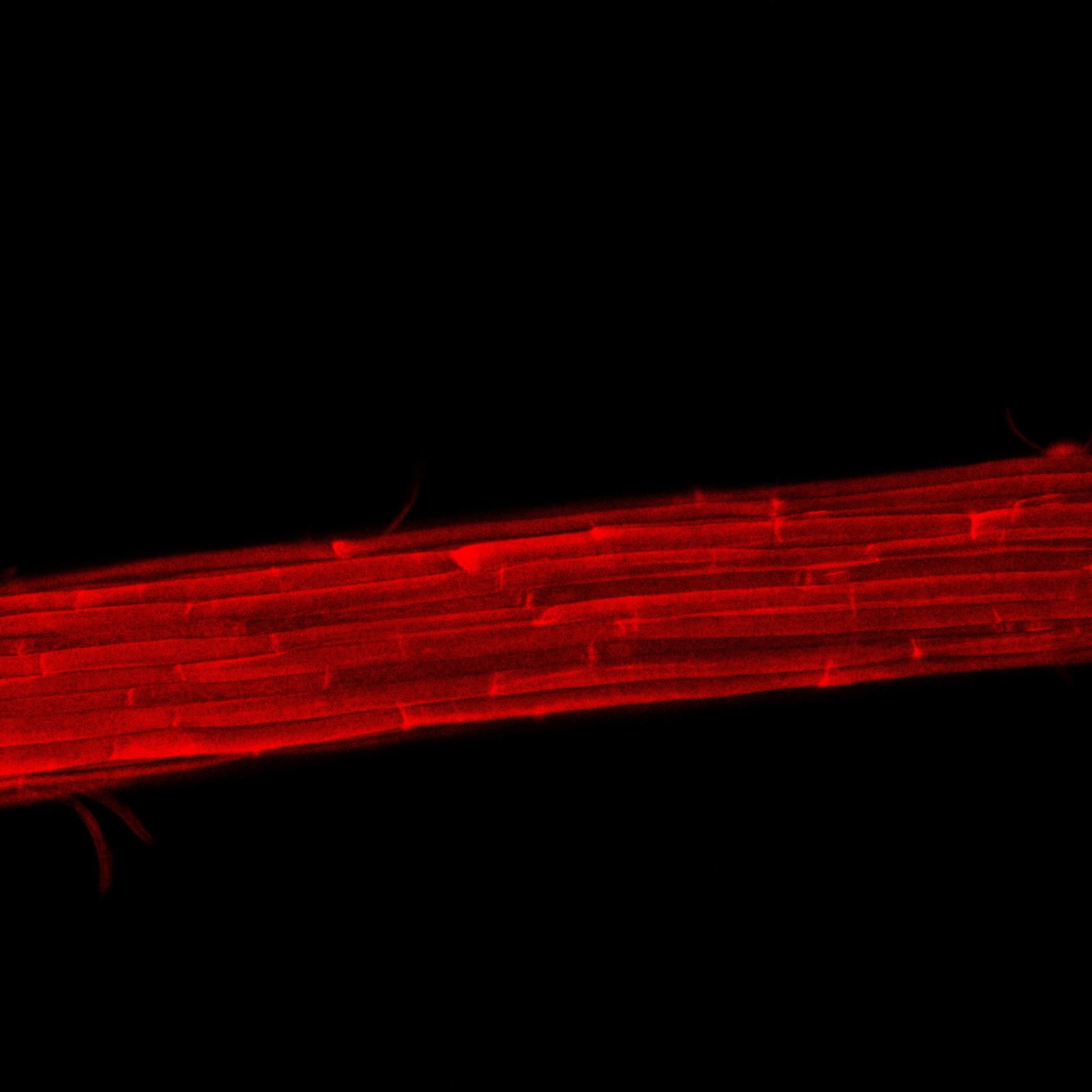
Figure 2. Congo red-stained root. It can be revealed that this image is what I used in last week’s post as a stand in for what I saw with the two-photon. Appearence in this region is similar. Sorry for the short cut!
The other breakthrough / puzzle piece / dropping shoe / was getting a copy of the virtual macro editor software for the LSM780 confocal. Actually, we don’t have it outright; instead, we have it as a demo for two months. Still, this means I can use the LSM780 and the gear (hardware and software) I brought according to plan.
Spurred on by the two-month constraint, I have set up a schedule to push through a dataset worth of roots. This coming week, I will calibrate the LSM780 by means of the stained celery xylem (a sample that has massive bands of cellulose whose orientation is known). Two days later, I will run sample roots thru to make sure actual root imaging goes well. Ever the optimist, I have planned out for the subsequent four weeks a schedule where I do three roots on each of Tuesday, Wednesday, and Thursday. At the end of all that, I’ll have 12 roots of each genotype. Let the show begin!